Hip (Trochanteric) Bursa Injections

Trochanteric bursa injections: targeted relief for hip pain

Trochanteric bursa injections are a highly effective treatment option for managing hip pain caused by inflammation in the bursa, which is a fluid-filled sac that cushions and reduces friction between bones, tendons, and muscles around the joint. This procedure involves the injection of medication directly into the bursa to alleviate pain, reduce swelling, and improve joint mobility.

What is trochanteric bursitis?

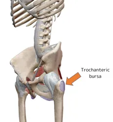
Trochanteric bursitis is a condition characterised by pain and inflammation of the trochanteric bursa, a fluid-filled sac located over the greater trochanter (the bony prominence on the outer side of the hip). Bursae are found throughout the body, positioned between bones and tendons to minimise friction during movement.

What are the causes of trochanteric bursitis?

Trochanteric bursitis can result from various factors, including:

  • Sports activities, such as football and soccer, that involve extensive running and overuse of the hip joint
  • Hip injuries from falls or impacts
  • Bone spurs in the hip or thighbone
  • Poor posture
  • Leg-length discrepancies
  • Complications from hip surgery
  • Medical conditions such as rheumatoid arthritis, gout, psoriasis, or thyroid disorders

What are the symptoms of trochanteric bursitis?

Common symptoms include:

  • Pain on the outer side of the hip, worsening with prolonged walking or stair climbing
  • Discomfort when lying on the affected side
  • Tenderness in the hip area
  • Stiffness in the hip joint
  • In severe cases, redness and swelling around the affected area

How is trochanteric bursitis diagnosed?

Diagnosis involves a review of your medical history, a physical examination of the hip, and sometimes imaging tests such as X-rays, MRI, or ultrasound.

In some cases, an anaesthetic injection may be used to confirm the diagnosis.


What is the treatment for trochanteric bursitis?

Conservative treatments are often effective and include:

  • Rest, ice application, and over-the-counter pain relievers (e.g.  anti-inflammatory gels)
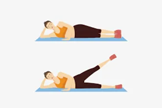
  • Physiotherapy:  The key is to strengthen the core muscles around the hip joint (the hip abductor muscles).

If symptoms persist, additional treatments may be required:

  • Trochanteric bursa steroid injections can help reduce inflammation and allow physiotherapy.
  • Extracorporeal shock wave therapy:  a non-invasive treatment that uses shock waves to treat hip bursitis.
  • Surgery: In severe cases, surgical removal of the bursa may be necessary

What are trochanteric bursa injections?

Trochanteric bursa injections involve injecting anti-inflammatory medication, typically corticosteroids, into the hip bursa. The bursa can become inflamed due to overuse, trauma, or underlying conditions like bursitis or arthritis, leading to significant pain and discomfort. By targeting the source of inflammation, hip bursa injections provide fast, targeted relief.

If you have persistent hip pain that limits your daily activities or if other conservative treatments have not provided adequate relief, hip bursa injections may be a suitable option. Consult with a specialist to evaluate your condition and discuss the potential benefits of this targeted pain relief method.

What are the advantages of these injections?
  • Targeted pain management: Direct relief to the inflamed area.
  • Quick procedure: Typically completed in a short visit.
  • Minimal downtime: Most patients can return to light activities soon after.
  • Non-surgical treatment: A less invasive option for managing pain.
  • Long-lasting relief: Effects can last weeks to months, depending on the patient but unfortunately time duration cannot be guaranteed.

What does the procedure involve?
  • Preparation: The patient is positioned comfortably, and the skin over the hip is cleaned with an antiseptic agent
  • Imaging guidance: Ultrasound or fluoroscopy is sometimes used to locate the exact position of the bursa.
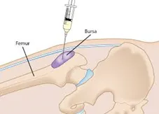
  • Injection: A thin needle is inserted into the bursa, and the medication is administered.
  • Completion: The needle is removed, and the area is cleaned and bandaged.

What is the post injection care?
  • Initial recovery: Patients may experience mild discomfort at the injection site, which usually resolves within a few days.
  • Activity recommendations: Light activities are encouraged, while intense physical activity should be avoided for 24-48 hours.
  • Follow-up care: Monitoring for changes in pain levels and any potential side effects.  Your surgeon will arrange follow-up with you and often at 6 weeks following the procedure.

What are the risks?

While hip bursa injections are generally safe, some of the potential risks include:

  • Temporary pain: Mild soreness at the injection site.
  • Skin reactions: Redness or slight swelling.
  • Infection (rare): Proper sterile techniques minimise this risk.
  • Allergic reactions: Rare but possible to medication components.

How can you prevent trochanteric bursitis?

To lower the risk of developing bursitis:

  • Maintain a healthy weight
  • Engage in regular exercise, ensuring proper form, warm-up, and stretching
  • Avoid activities that excessively strain the hips
  • Use orthotic devices to correct leg-length discrepancies
  • Strengthen and maintain flexibility in the hip muscles

Summary: precision pain relief for hip discomfort

Hip (trochanteric) bursa injections offer a highly effective solution for managing hip pain and inflammation. With minimal downtime and a targeted approach, these injections can help improve quality of life, allowing patients to return to their daily activities with reduced pain and enhanced joint function. Contact a specialist to see if hip bursa injections are right for you and take a step toward better hip health.

Physiotherapy

Knee physiotherapy protocols following knee replacement surgery

Physiotherapy is an essential part of recovery after knee replacement surgery. It helps restore mobility, improve strength, and ensure the best long-term outcomes. Below is a general outline of knee physiotherapy protocols, which may be tailored to each patient’s specific needs by their healthcare provider:

1. Immediate Postoperative Phase (Days 1–7)

Goals:

  • Prevent complications like blood clots or stiffness.
  • Begin gentle movement to restore range of motion.

Activities:

  • Ankle Pumps: To promote blood circulation.
  • Quad Sets: Tighten thigh muscles to prevent muscle atrophy.
  • Passive Range of Motion: Assisted bending and straightening of the knee.
  • Walking with Assistance: Using a walker or crutches for short distances.
2. Early Recovery Phase (Weeks 2–6)

Goals:

  • Increase range of motion (aim for 90–120 degrees of knee flexion).
  • Improve weight-bearing ability.
  • Strengthen surrounding muscles.

Activities:

  • Straight Leg Raises: Strengthen quadriceps without stressing the joint.
  • Heel Slides: Gradual bending of the knee while lying down.
  • Stationary Cycling: Low resistance to enhance mobility.
  • Step-Ups: Gentle step exercises on a low platform.
3. Intermediate Phase (Weeks 6–12)

Goals:

  • Normalize walking patterns.
  • Enhance muscle strength and joint stability.

Activities:

  • Balance Exercises: Use a stability board or single-leg stance.
  • Resistance Training: Light weights or resistance bands for leg muscles.
  • Walking Without Support: Gradually increase distances.
  • Functional Movements: Gentle squats and lunges to prepare for daily activities.
4. Advanced Phase (Months 3–6)

Goals:

  • Achieve full range of motion.
  • Return to low-impact activities.

Activities:

  • Higher Resistance Training: Focus on strengthening quads, hamstrings, and calves.
  • Advanced Balance Training: Progressively challenging exercises to improve coordination.
  • Low-Impact Activities: Swimming, walking, or cycling for cardiovascular fitness.
5. Maintenance Phase (Beyond 6 Months)

Goals:

  • Maintain strength and mobility.
  • Safely return to routine activities or sports, as advised.

Activities:

  • Continue with strengthening and mobility exercises.
  • Low-impact sports and recreational activities.
  • Periodic follow-ups with a physiotherapist for guidance.
Key Points to Remember:
  • Consistency is crucial for optimal recovery.
  • Avoid high-impact activities that may stress the joint.
  • Always follow your surgeon’s and physiotherapist’s advice regarding activity progression.
Hip physiotherapy protocols following hip replacement surgery

Physiotherapy after hip replacement surgery plays a vital role in regaining mobility, strengthening muscles, and ensuring the longevity of the new joint. Below is an overview of the typical physiotherapy protocols, divided into phases for a structured recovery process:

1. Immediate Postoperative Phase (Days 1–7)

Goals:

  • Prevent complications like blood clots or stiffness.
  • Begin gentle movement and weight-bearing exercises.

Activities:

  • Ankle Pumps: To improve blood circulation and reduce swelling.
  • Quad Sets and Glute Squeezes: Strengthen thigh and hip muscles.
  • Heel Slides: Gradual bending of the hip and knee while lying down.
  • Assisted Walking: Use of a walker or crutches for short distances.
2. Early Recovery Phase (Weeks 2–6)

Goals:

  • Improve range of motion and muscle strength.
  • Transition to more independent mobility.

Activities:

  • Straight Leg Raises: Strengthen quadriceps without stressing the hip.
  • Hip Abductions: Lifting the leg sideways while lying down or standing.
  • Stationary Cycling: Gentle cycling with low resistance.
  • Weight-Bearing Exercises: Gradual increase in walking distances with support.
3. Intermediate Recovery Phase (Weeks 6–12)

Goals:

  • Normalize walking patterns and restore functional movement.
  • Strengthen surrounding muscles and improve joint stability.

Activities:

  • Step-Ups: Controlled step exercises on a low platform.
  • Balance Training: Single-leg stance exercises with support if needed.
  • Resistance Band Workouts: Strengthen hip abductors and extensors.
  • Walking Without Support: Gradually increase distances, focusing on proper gait.
4. Advanced Recovery Phase (Months 3–6)

Goals:

  • Build strength and flexibility to support routine activities.
  • Begin low-impact recreational activities.

Activities:

  • Advanced Strengthening Exercises: Light weights for leg muscles.
  • Functional Training: Gentle squats and lunges to prepare for daily activities.
  • Low-Impact Aerobic Activities: Swimming, cycling, or walking for endurance.
5. Maintenance Phase (Beyond 6 Months)

Goals:

  • Maintain strength and mobility.
  • Safely resume hobbies and low-impact sports.

Activities:

  • Continue strengthening exercises and stretching routines.
  • Engage in low-impact activities like yoga or Pilates.
  • Follow up with your physiotherapist periodically to ensure progress.
Important Tips:
  • Avoid high-impact activities or heavy lifting during the early phases.
  • Adhere to hip precautions (e.g., avoiding excessive bending or twisting) as advised by your surgeon.
  • Pain is a signal to stop—don’t push through discomfort.
  • Stay consistent with your physiotherapy exercises for optimal recovery.
Knee physiotherapy protocols following knee meniscal surgery (meniscus repaired vs trimmed (partial meniscectomy)
1. Post-Operative Phase: (Weeks 1-2)

Goal: Control pain, swelling, and inflammation, protect the surgical site, and initiate early range of motion (ROM).

  • Meniscal Repair Protocol:
    • Weight-bearing: Non-weight bearing (NWB) or partial weight-bearing (PWB) with crutches for 4-6 weeks to protect the repair.
    • ROM: Passive and active-assisted range of motion exercises within pain tolerance.
    • Exercise: Isometric quadriceps contractions, ankle pumps, and straight leg raises to avoid muscle atrophy.
    • Protection: Knee brace with limited flexion (0-90 degrees), as directed by the surgeon.
  • Partial Meniscectomy Protocol:
    • Weight-bearing: Full weight-bearing (FWB) allowed immediately or after a few days depending on the surgeon's preference.
    • ROM: Gentle range of motion exercises, avoiding deep squats or high-load activities.
    • Exercise: Focus on quadriceps strengthening, and gentle hamstring and calf stretches.
    • Protection: No bracing is generally needed, but the patient should avoid twisting or pivoting.
2. Early Rehabilitation Phase: (Weeks 3-6)

Goal: Improve ROM, decrease swelling, and initiate strength training.

  • Meniscal Repair Protocol:
    • Weight-bearing: Gradual increase in weight-bearing, progressing to full weight-bearing by 6 weeks if tolerated.
    • ROM: Continue working on achieving full extension and 90-110 degrees of flexion.
    • Strengthening: Quadriceps and hamstring strengthening exercises, focusing on pain-free motion.
    • Exercise: Progress to closed-chain exercises like leg presses (avoiding deep knee bends initially), hip abduction, and mini squats.
  • Partial Meniscectomy Protocol:
    • Weight-bearing: Full weight-bearing is maintained.
    • ROM: Full knee extension and flexion should be encouraged.
    • Strengthening: Quadriceps and hamstring strengthening with light resistance bands or weights.
    • Exercise: Begin low-impact closed-chain exercises, including stationary cycling and pool therapy if available.
3. Intermediate Rehabilitation Phase: (Weeks 7-12)

Goal: Build strength, improve functional mobility, and gradually return to more dynamic activities.

  • Meniscal Repair Protocol:
    • Strengthening: Progressive resistance training (leg press, squats) and proprioceptive exercises (e.g., balance and stability work).
    • Activity: Begin low-impact activities such as cycling and swimming, avoid running or jumping.
    • Functional Training: Begin agility drills and proprioceptive training.
  • Partial Meniscectomy Protocol:
    • Strengthening: Continue strengthening exercises with added resistance.
    • Activity: Running can begin if there are no significant signs of pain or swelling.
    • Functional Training: Return to light activities, with controlled motion, agility drills, and functional tasks.
4. Advanced Rehabilitation Phase: (Weeks 13-24)

Goal: Continue to strengthen the knee, return to normal function, and resume sports and recreational activities.

  • Meniscal Repair Protocol:
    • Strengthening: Continue strengthening exercises (focus on quads, hamstrings, and calves) and increase resistance or weights as tolerated.
    • Activity: Low-impact activities like swimming or cycling. Running may be introduced cautiously at 12-16 weeks, depending on the surgeon's approval.
    • Return to Sports: Typically after 6 months, depending on healing progress, sports-specific exercises may begin.
  • Partial Meniscectomy Protocol:
    • Strengthening: Full return to weight-bearing exercises with more advanced resistance.
    • Activity: Gradual progression of running, plyometrics, and sport-specific drills.
    • Return to Sports: Return to full activity and sports after 12-16 weeks if cleared by the surgeon.
5. Long-Term Maintenance Phase: (Months 6+)

Goal: Maintain knee strength and function and prevent further injury.

  • Meniscal Repair Protocol:
    • Full return to sports may take 6-12 months or more, depending on the type and success of the meniscal repair.
    • Continued strengthening and conditioning exercises.
  • Partial Meniscectomy Protocol:
    • Return to normal activity should be well-established by 6 months.
    • Ongoing strengthening exercises and proprioceptive training to ensure knee stability.
Key Differences:
  • Weight-bearing restrictions: More stringent in meniscal repair versus partial meniscectomy.
  • Return to activity: Generally, patients following meniscal repair take longer to return to sports due to the need for healing of the repaired meniscus.
  • Intensity of exercise: Those with a meniscal repair often progress more slowly in terms of weight-bearing and exercise intensity.

It is crucial for patients to follow these protocols closely, with regular follow-up appointments with their surgeon or physiotherapist to ensure proper healing and recovery progress.

Physiotherapy to manage hip pain from osteoarthritis conservatively

Managing hip pain from osteoarthritis (OA) conservatively through physiotherapy involves a combination of exercises, manual therapy, and lifestyle modifications aimed at reducing pain, improving function, and preventing further deterioration of the joint. Here's a detailed approach to physiotherapy for managing hip pain caused by OA

1.Pain Management
  • Heat/Cold Therapy: Use cold packs to reduce inflammation and heat to relax muscles.
  • TENS Therapy: Electrical stimulation can help manage pain.
2. Strengthening Exercises
  • Glutes and Quads: Exercises like leg raises, squats, and bridges help stabilize the hip joint.
  • Hamstrings: Strengthening the hamstrings to balance leg muscles and reduce stress on the hip.
3. Range of Motion (ROM) Exercises
  • Hip Flexion/Extension Stretching: Gentle stretches to improve flexibility and reduce stiffness.
  • Rotator Stretching: Helps improve internal and external hip rotation.
4. Manual Therapy
  • Joint Mobilization: Gentle, hands-on techniques to improve joint movement.
  • Soft Tissue Mobilization: Focus on relieving tightness in surrounding muscles like the hip flexors and glutes.
5. Posture and Gait Training
  • Posture Awareness: Educate on proper body mechanics to avoid excess strain on the hip joint.
  • Gait Training: Adjust walking techniques to reduce impact on the hip.
6. Core Stability and Balance
  • Core Strengthening: Exercises like planks to improve posture and reduce hip strain.
  • Balance Training: Simple exercises like single-leg stands to improve stability.
7. Aquatic Therapy
  • Water Exercises: Buoyancy reduces joint stress, making it easier to perform strengthening and stretching exercises.
8. Long-Term Maintenance
  • Home Exercise Program: Provide simple exercises for patients to do at home.
  • Gradual Activity Increase: As strength and mobility improve, encourage more activity like swimming and walking.
physiotherapy to manage hip pain from trochanteric hip bursitis

Trochanteric hip bursitis causes pain on the outer side of the hip due to inflammation of the bursa. Physiotherapy can help reduce pain, improve function, and prevent recurrence.

1. Pain Management
  • Ice Therapy: Apply ice to the outer hip for 15-20 minutes, several times a day to reduce inflammation and pain.
  • Heat Therapy: Use heat to relax tight muscles once acute pain subsides.
  • TENS Therapy: Electrical nerve stimulation can assist with pain relief.
2. Stretching Exercises
  • Hip Flexor Stretch: Relieves tightness in the front of the hip.
  • IT Band Stretch: Reduces tension on the outer hip.
  • Glute Stretch: Helps ease tightness in the glutes and surrounding muscles.
3. Strengthening Exercises
  • Gluteus Medius Strengthening: Side-lying leg lifts and clamshells target the gluteus medius to support the hip.
  • Hip Abduction: Strengthens the hip abductors to stabilize the joint and reduce stress on the bursa.
  • Core Strengthening: Bridges and planks improve pelvic stability and reduce strain on the hip.
4. Manual Therapy
  • Soft Tissue Mobilization: Gentle massage can relieve muscle tightness around the hip.
  • Joint Mobilizations: Physiotherapists may use manual techniques to enhance hip mobility.
5. Posture and Gait Training
  • Postural Awareness: Correct standing, sitting, and walking positions reduce hip strain.
  • Gait Training: Proper walking mechanics can prevent aggravation of the bursitis.
6. Balance and Stability Training
  • Balance Exercises: Single-leg stands improve stability and reduce compensatory movements.
  • Functional Exercises: Gradually incorporate exercises that mimic daily activities to strengthen the hip.
7. Gradual Return to Activity
  • Low-Impact Activities: Start with swimming or cycling to strengthen without stressing the hip.
  • Avoid Overuse: Gradually increase activity levels to prevent flare-ups.

This conservative approach can significantly reduce pain and improve hip function in individuals with trochanteric hip bursitis.

Physiotherapy to manage knee pain from osteoarthritis conservatively

Physiotherapy plays a crucial role in managing knee pain from osteoarthritis (OA) conservatively by focusing on pain relief, improving joint function, and preventing further damage. Here's a concise breakdown of the approach:

1. Pain Management
  • Ice Therapy: Apply ice to the knee for 15-20 minutes to reduce inflammation and pain.
  • Heat Therapy: Use heat to relax muscles around the knee after acute pain subsides.
  • TENS Therapy: Transcutaneous electrical nerve stimulation can provide pain relief by stimulating nerves.
2. Strengthening Exercises
  • Quadriceps Strengthening: Exercises like straight leg raises and squats help strengthen the quadriceps, which support the knee joint.
  • Hamstring Strengthening: Strengthening the hamstrings reduces strain on the knee and balances the muscles around the joint.
  • Glute Strengthening: Targeting the glutes with exercises like bridges helps improve pelvic stability and reduce knee strain.
3. Range of Motion (ROM) Exercises
  • Knee Flexion/Extension: Gentle stretches to improve the knee's ability to bend and straighten.
  • Heel and Toe Raises: To maintain mobility and reduce stiffness in the knee joint.
4. Manual Therapy
  • Joint Mobilization: Hands-on techniques from the physiotherapist can improve knee mobility and alleviate discomfort.
  • Soft Tissue Mobilization: Massage or myofascial release to ease muscle tightness and improve blood flow around the knee.
5. Posture and Gait Training
  • Postural Awareness: Correct alignment when standing, sitting, and walking can help reduce stress on the knee.
  • Gait Training: Learning to walk with a more efficient stride can alleviate pressure on the knee joint.
6. Balance and Stability Training
  • Balance Exercises: Single-leg stands or balance boards can improve knee stability and prevent falls.
  • Proprioception Training: Exercises that help improve the knee’s sense of position can reduce the risk of injury.
7. Aquatic Therapy
  • Water-Based Exercises: Swimming or water aerobics can reduce impact on the knee while strengthening muscles and improving range of motion.
8. Gradual Return to Activity
  • Low-Impact Activities: Activities like cycling or walking can be introduced to build strength without overloading the knee.
  • Avoid Overuse: Gradually increasing activity levels ensures the knee is not overstrained.

This conservative physiotherapy approach aims to improve knee function, reduce pain, and help patients manage osteoarthritis effectively.