Hip Replacement

What is hip replacement surgery?

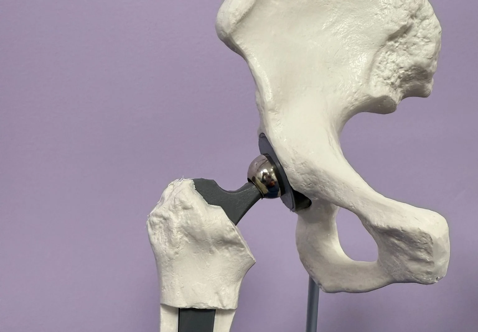
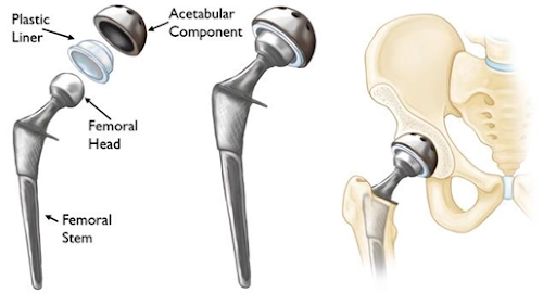
Hip replacement, or hip arthroplasty, is a surgical procedure where the damaged portions of the hip joint are replaced with artificial components. These prosthetics mimic the natural movement of the hip, allowing patients to regain pain-free mobility.

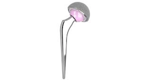
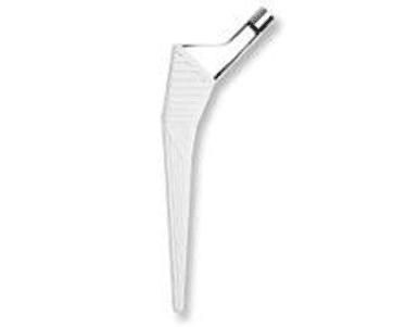
  • The damaged femoral head is removed and replaced with a metal stem, which is inserted into the hollow center of the femur. The stem can be secured using either cement or a “press-fit” technique.
  • A metal or ceramic ball is attached to the upper part of the stem, replacing the removed femoral head.
  • The damaged cartilage lining the socket (acetabulum) is also removed and replaced with a metal socket, which may be secured using screws or cement.
  • To ensure smooth movement, a spacer made of plastic, ceramic, or metal (called a liner) is placed between the new ball and socket.
What are the types of hip replacement procedures?
  • Total hip replacement: Both the femoral head (ball) and acetabulum (socket) are replaced.
  • Partial hip replacement: Only the femoral head is replaced, often for specific types of fractures.
  • Hip resurfacing replacement:  This type of hip replacement involves resurfacing the hip joint rather than fully replacing it. It is a bone-preserving procedure designed to alleviate hip pain caused by arthritis or other conditions.
  • Minimally invasive hip replacement: Smaller incisions and advanced techniques for reduced recovery time and scarring.
  • Revision hip replacement: Replacement of a previously implanted hip prosthesis that has worn out or failed.

How does total hip replacement surgery allow mobility and comfort?

Hip replacement surgery is a transformative procedure designed to alleviate pain, improve mobility, and enhance quality of life for individuals suffering from debilitating hip joint conditions. This advanced surgical option replaces a damaged or worn-out hip joint with a prosthetic implant, restoring function and comfort.

The hip typically moves smoothly because the ends of the bones are covered with a protective layer of tough tissue known as cartilage. When the cartilage is worn down or damaged, often due to injury, the bones begin to rub against each other, leading to pain and stiffness in the joint.

In a hip replacement, the damaged components of the hip are replaced with metal, ceramic, and plastic parts.

Who can benefit from hip replacement?

A hip replacement may be necessary if one or both of your hip joints become damaged, leading to persistent pain or difficulty with everyday activities such as walking, driving, or getting dressed.

It is most often used to relieve pain and stiffness in the hip joint caused by osteoarthritis.

Common causes of hip joint damage include:

  • Osteoarthritis: Also known as “wear-and-tear arthritis,” where the protective cartilage cushioning the ends of bones within a hip joint wears away with time, causing the bones to rub against each other.  This results in pain, stiffness, swelling, and reduced mobility in the affected joint. It is the most common type of arthritis and frequently affects weight-bearing joints like the knees, hips, and spine.
  • Rheumatoid arthritis: An autoimmune condition where the immune system mistakenly attacks the joint lining, resulting in pain and stiffness.
  • Psoriatic arthritis:  A form of arthritis that is linked to psoriasis- a chronic skin condition.
  • Hip fracture: Severe damage to the hip joint from a fall or similar accident may require replacement.


Other health conditions and issues that can damage your hip joint enough to require a hip replacement include:

  • Avascular necrosis: a painful bone condition that can deteriorate with time caused by reduced blood supply to one of your bones.  A variety of risk factors for this include a traumatic event, sickle cell disease, steroid use, alcoholism, autoimmune disorders, and hypercoagulable states.
  • Femoroacetabular impingement syndrome: a condition where the hip joint’s bones are abnormally shaped causing them to rub together leading to pain and discomfort. 
  • Hip Dysplasia:  a condition where the hip socket does not fully cover the ball portion of the upper thigh bone (femur) at the hip joint.  This can cause the hip socket to be too shallow, or the ball of the hip socket to be loose or dislocated.
  • Tumours:  a group of abnormal cells that form lumps or growths which can start in any cells in the body and behave differently if they are cancerous (malignant) or benign (non-cancerous).
  • Perthes disease:  It affects the growing end of the femur bone that fits into your child’s hip socket and presents in childhood.  It can cause the end of the bone to break down and then regenerate over several years.

What are the other common causes of pain in the hip region?
  • Hip bursitis – Inflammation of the bursae, small fluid-filled sacs that cushion the hip joint.
  • Tendonitis – Inflammation of tendons around the hip due to overuse or repetitive strain.
  • Hip labral tears – Damage to the cartilage surrounding the hip socket, often from injury or repetitive motion.
  • Muscle strains – Overstretching or tearing of muscles around the hip from physical activity.
  • Sciatica – Pain radiating from the lower back to the hip due to compression of the sciatic nerve.

What are the benefits of hip replacement surgery?
  • Pain Relief: Significant reduction or elimination of hip pain.
  • Improved Mobility: Greater range of motion and ability to perform daily activities.
  • Enhanced Quality of Life: Increased independence and participation in recreational activities.
  • Durable Results: Modern implants often can last 15-20 years or more.

What are the alternatives to hip replacement surgery?

Most hip pain issues typically do not require surgery and can be managed with non-invasive treatments, including

  • pain medication
  • walking aids
  • physiotherapy
  • joint injections.

What happens during a hip replacement?

If you’re undergoing a hip replacement, you’ll typically be admitted to the hospital on the day of your surgery.

The procedure involves either a spinal anaesthetic, which numbs the body from the waist down while keeping you awake, or a general anaesthetic, which puts you to sleep throughout the operation.

If you opt for a spinal anaesthetic, you may also be given a sedative to help you stay calm and drowsy.

The surgery generally lasts around 1 to 2 hours, and the specific approach taken will depend on factors such as the extent of hip damage, its underlying cause, and your overall health.

The procedure involves the following steps:

  • Anaesthesia: Administered to ensure comfort during surgery.
  • Joint access: An incision is made to access the hip joint.
  • Damaged tissue removal: The damaged cartilage and bone are removed.
  • Prosthesis placement: The artificial components (metal, ceramic, or plastic) are securely placed to form a new joint.
  • Closure: The incision is closed, and the area is bandaged.

What does the recovery and rehabilitation involve?
  • Hospital stay: Patients typically stay in the hospital for 0-3 days. 
  • Pain management: Medications and physical therapy help manage postoperative pain.
  • Rehabilitation: Exercises to strengthen muscles and improve joint movement start shortly after surgery.
  • Return to activity: Most individuals resume normal activities within 12 weeks, with improvements continuing over several months.  Improvement in pain have been shown to occur up to 1- 1.5 years following the surgery.

What are the potential risks and complications?

Your surgeon takes every precaution to minimise the risks of surgery, using state-of-the-art techniques and providing comprehensive preoperative and postoperative care.

Rest assured, your consultant will carefully assess the risks and benefits before recommending surgery.

During your initial consultation, they will also discuss any specific risks relevant to your situation.

While hip replacement is a widely successful procedure, risks are rare but possible and some of these can include:

  • Infection.
  • Nerve or blood vessel damage
  • Blood clots.
  • Implant wear or loosening over time.
  • Dislocation of the prosthetic joint.
  • Altered leg length
  • Bone fracture
  • Bone forming in muscles around the replacement
  • Anaesthetic risk

What is the recovery like at home?  Tips for a smooth recovery

When you return home, you may feel quite tired. Prioritise rest initially, then gradually increase your activity levels to aid recovery.

To strengthen your hip, protect it from further injury, and support wound healing, follow these guidelines:

  • Perform the exercises recommended by your physiotherapist 3 to 4 times daily for 6 weeks. Listen to your body and do not worry if cannot keep up as recommended.
  • Engage in low-impact activities like walking and gradually increase the distance as you feel comfortable.  The balance between strengthening exercises versus walking distance should favour strengthening exercises initially, as this will allow you to achieve an increased walking distance.
  • Avoid bending your hip beyond a 90-degree angle.
  • Try to sleep on your back for 6-8 weeks (If unable, one can sleep on the side, using a pillow between legs, with the operative side facing upwards)
  • Avoid significant twisting movements of the hip and crossing your legs.
  • Refrain from lifting heavy objects or engaging in strenuous activities.
  • Continue using crutches or a walking stick for support as long as needed.
  • If you experience discomfort, take the prescribed painkillers until the pain subsides.
  • Opt for showers instead of baths to prevent strain on your hip.
  • It is critically important to keep the wound dry during healing which can take up to 3 weeks and so protect it when having a shower despite the dressing.


These steps will help ensure a safe and effective recovery.


How to maintain a healthy hip after surgery?
  • Follow rehabilitation guidelines: Adhere to physical therapy and exercise routines.
  • Prevent falls: Use assistive devices and take precautions to avoid accidents.
  • Healthy lifestyle: Maintain a balanced diet and healthy weight to reduce stress on the joint.
  • Regular check-ups: Monitor the condition of the prosthesis through routine medical evaluations.

When can I drive a car after surgery?

You may be able to drive again approximately six weeks after your surgery, although this timeframe can vary between individuals. Your surgeon will provide specific guidance on when you’re ready. Do not drive until you feel confident in controlling your vehicle and always confirm with both your surgeon and your insurance provider before resuming driving.


When can I return to work after surgery?

Before going back to work, consult your doctor for personalised advice. As a general guideline:

  • Sedentary jobs (e.g. office-based work): You can typically return after 4–6 weeks.
  • Mixed-labour jobs (e.g. teacher): You may return after about 3 months.
  • Manual labour jobs (e.g. builder): You should wait approximately 6 months before resuming work.

How soon can you fly after hip replacement surgery?

Most surgeons and airlines recommend you should not fly within 3 months of surgery because of the increased risk of developing blood clots (Deep vein thrombosis, pulmonary embolism).

Please check with your surgeon for any advice regarding air travel and any precautions to take if you have additional risk factors for blood clots.

What is the follow-up schedule after surgery?

A wound check will be required at approximately 14 days after your surgery with a healthcare professional.

A routine follow-up appointment with your surgeon will be needed around 6–8 weeks after your surgery.


Summary: Embrace a pain-free future

Hip replacement surgery offers a reliable solution for individuals living with chronic hip pain and limited mobility. With advanced techniques and high success rates, this procedure can restore your freedom of movement and significantly enhance your quality of life. If you’re considering hip replacement, schedule a consultation to explore your options and embark on the journey to a pain-free life.

Physiotherapy

Knee physiotherapy protocols following knee replacement surgery

Physiotherapy is an essential part of recovery after knee replacement surgery. It helps restore mobility, improve strength, and ensure the best long-term outcomes. Below is a general outline of knee physiotherapy protocols, which may be tailored to each patient’s specific needs by their healthcare provider:

1. Immediate Postoperative Phase (Days 1–7)

Goals:

  • Prevent complications like blood clots or stiffness.
  • Begin gentle movement to restore range of motion.

Activities:

  • Ankle Pumps: To promote blood circulation.
  • Quad Sets: Tighten thigh muscles to prevent muscle atrophy.
  • Passive Range of Motion: Assisted bending and straightening of the knee.
  • Walking with Assistance: Using a walker or crutches for short distances.
2. Early Recovery Phase (Weeks 2–6)

Goals:

  • Increase range of motion (aim for 90–120 degrees of knee flexion).
  • Improve weight-bearing ability.
  • Strengthen surrounding muscles.

Activities:

  • Straight Leg Raises: Strengthen quadriceps without stressing the joint.
  • Heel Slides: Gradual bending of the knee while lying down.
  • Stationary Cycling: Low resistance to enhance mobility.
  • Step-Ups: Gentle step exercises on a low platform.
3. Intermediate Phase (Weeks 6–12)

Goals:

  • Normalize walking patterns.
  • Enhance muscle strength and joint stability.

Activities:

  • Balance Exercises: Use a stability board or single-leg stance.
  • Resistance Training: Light weights or resistance bands for leg muscles.
  • Walking Without Support: Gradually increase distances.
  • Functional Movements: Gentle squats and lunges to prepare for daily activities.
4. Advanced Phase (Months 3–6)

Goals:

  • Achieve full range of motion.
  • Return to low-impact activities.

Activities:

  • Higher Resistance Training: Focus on strengthening quads, hamstrings, and calves.
  • Advanced Balance Training: Progressively challenging exercises to improve coordination.
  • Low-Impact Activities: Swimming, walking, or cycling for cardiovascular fitness.
5. Maintenance Phase (Beyond 6 Months)

Goals:

  • Maintain strength and mobility.
  • Safely return to routine activities or sports, as advised.

Activities:

  • Continue with strengthening and mobility exercises.
  • Low-impact sports and recreational activities.
  • Periodic follow-ups with a physiotherapist for guidance.
Key Points to Remember:
  • Consistency is crucial for optimal recovery.
  • Avoid high-impact activities that may stress the joint.
  • Always follow your surgeon’s and physiotherapist’s advice regarding activity progression.
Hip physiotherapy protocols following hip replacement surgery

Physiotherapy after hip replacement surgery plays a vital role in regaining mobility, strengthening muscles, and ensuring the longevity of the new joint. Below is an overview of the typical physiotherapy protocols, divided into phases for a structured recovery process:

1. Immediate Postoperative Phase (Days 1–7)

Goals:

  • Prevent complications like blood clots or stiffness.
  • Begin gentle movement and weight-bearing exercises.

Activities:

  • Ankle Pumps: To improve blood circulation and reduce swelling.
  • Quad Sets and Glute Squeezes: Strengthen thigh and hip muscles.
  • Heel Slides: Gradual bending of the hip and knee while lying down.
  • Assisted Walking: Use of a walker or crutches for short distances.
2. Early Recovery Phase (Weeks 2–6)

Goals:

  • Improve range of motion and muscle strength.
  • Transition to more independent mobility.

Activities:

  • Straight Leg Raises: Strengthen quadriceps without stressing the hip.
  • Hip Abductions: Lifting the leg sideways while lying down or standing.
  • Stationary Cycling: Gentle cycling with low resistance.
  • Weight-Bearing Exercises: Gradual increase in walking distances with support.
3. Intermediate Recovery Phase (Weeks 6–12)

Goals:

  • Normalize walking patterns and restore functional movement.
  • Strengthen surrounding muscles and improve joint stability.

Activities:

  • Step-Ups: Controlled step exercises on a low platform.
  • Balance Training: Single-leg stance exercises with support if needed.
  • Resistance Band Workouts: Strengthen hip abductors and extensors.
  • Walking Without Support: Gradually increase distances, focusing on proper gait.
4. Advanced Recovery Phase (Months 3–6)

Goals:

  • Build strength and flexibility to support routine activities.
  • Begin low-impact recreational activities.

Activities:

  • Advanced Strengthening Exercises: Light weights for leg muscles.
  • Functional Training: Gentle squats and lunges to prepare for daily activities.
  • Low-Impact Aerobic Activities: Swimming, cycling, or walking for endurance.
5. Maintenance Phase (Beyond 6 Months)

Goals:

  • Maintain strength and mobility.
  • Safely resume hobbies and low-impact sports.

Activities:

  • Continue strengthening exercises and stretching routines.
  • Engage in low-impact activities like yoga or Pilates.
  • Follow up with your physiotherapist periodically to ensure progress.
Important Tips:
  • Avoid high-impact activities or heavy lifting during the early phases.
  • Adhere to hip precautions (e.g., avoiding excessive bending or twisting) as advised by your surgeon.
  • Pain is a signal to stop—don’t push through discomfort.
  • Stay consistent with your physiotherapy exercises for optimal recovery.
Knee physiotherapy protocols following knee meniscal surgery (meniscus repaired vs trimmed (partial meniscectomy)
1. Post-Operative Phase: (Weeks 1-2)

Goal: Control pain, swelling, and inflammation, protect the surgical site, and initiate early range of motion (ROM).

  • Meniscal Repair Protocol:
    • Weight-bearing: Non-weight bearing (NWB) or partial weight-bearing (PWB) with crutches for 4-6 weeks to protect the repair.
    • ROM: Passive and active-assisted range of motion exercises within pain tolerance.
    • Exercise: Isometric quadriceps contractions, ankle pumps, and straight leg raises to avoid muscle atrophy.
    • Protection: Knee brace with limited flexion (0-90 degrees), as directed by the surgeon.
  • Partial Meniscectomy Protocol:
    • Weight-bearing: Full weight-bearing (FWB) allowed immediately or after a few days depending on the surgeon's preference.
    • ROM: Gentle range of motion exercises, avoiding deep squats or high-load activities.
    • Exercise: Focus on quadriceps strengthening, and gentle hamstring and calf stretches.
    • Protection: No bracing is generally needed, but the patient should avoid twisting or pivoting.
2. Early Rehabilitation Phase: (Weeks 3-6)

Goal: Improve ROM, decrease swelling, and initiate strength training.

  • Meniscal Repair Protocol:
    • Weight-bearing: Gradual increase in weight-bearing, progressing to full weight-bearing by 6 weeks if tolerated.
    • ROM: Continue working on achieving full extension and 90-110 degrees of flexion.
    • Strengthening: Quadriceps and hamstring strengthening exercises, focusing on pain-free motion.
    • Exercise: Progress to closed-chain exercises like leg presses (avoiding deep knee bends initially), hip abduction, and mini squats.
  • Partial Meniscectomy Protocol:
    • Weight-bearing: Full weight-bearing is maintained.
    • ROM: Full knee extension and flexion should be encouraged.
    • Strengthening: Quadriceps and hamstring strengthening with light resistance bands or weights.
    • Exercise: Begin low-impact closed-chain exercises, including stationary cycling and pool therapy if available.
3. Intermediate Rehabilitation Phase: (Weeks 7-12)

Goal: Build strength, improve functional mobility, and gradually return to more dynamic activities.

  • Meniscal Repair Protocol:
    • Strengthening: Progressive resistance training (leg press, squats) and proprioceptive exercises (e.g., balance and stability work).
    • Activity: Begin low-impact activities such as cycling and swimming, avoid running or jumping.
    • Functional Training: Begin agility drills and proprioceptive training.
  • Partial Meniscectomy Protocol:
    • Strengthening: Continue strengthening exercises with added resistance.
    • Activity: Running can begin if there are no significant signs of pain or swelling.
    • Functional Training: Return to light activities, with controlled motion, agility drills, and functional tasks.
4. Advanced Rehabilitation Phase: (Weeks 13-24)

Goal: Continue to strengthen the knee, return to normal function, and resume sports and recreational activities.

  • Meniscal Repair Protocol:
    • Strengthening: Continue strengthening exercises (focus on quads, hamstrings, and calves) and increase resistance or weights as tolerated.
    • Activity: Low-impact activities like swimming or cycling. Running may be introduced cautiously at 12-16 weeks, depending on the surgeon's approval.
    • Return to Sports: Typically after 6 months, depending on healing progress, sports-specific exercises may begin.
  • Partial Meniscectomy Protocol:
    • Strengthening: Full return to weight-bearing exercises with more advanced resistance.
    • Activity: Gradual progression of running, plyometrics, and sport-specific drills.
    • Return to Sports: Return to full activity and sports after 12-16 weeks if cleared by the surgeon.
5. Long-Term Maintenance Phase: (Months 6+)

Goal: Maintain knee strength and function and prevent further injury.

  • Meniscal Repair Protocol:
    • Full return to sports may take 6-12 months or more, depending on the type and success of the meniscal repair.
    • Continued strengthening and conditioning exercises.
  • Partial Meniscectomy Protocol:
    • Return to normal activity should be well-established by 6 months.
    • Ongoing strengthening exercises and proprioceptive training to ensure knee stability.
Key Differences:
  • Weight-bearing restrictions: More stringent in meniscal repair versus partial meniscectomy.
  • Return to activity: Generally, patients following meniscal repair take longer to return to sports due to the need for healing of the repaired meniscus.
  • Intensity of exercise: Those with a meniscal repair often progress more slowly in terms of weight-bearing and exercise intensity.

It is crucial for patients to follow these protocols closely, with regular follow-up appointments with their surgeon or physiotherapist to ensure proper healing and recovery progress.

Physiotherapy to manage hip pain from osteoarthritis conservatively

Managing hip pain from osteoarthritis (OA) conservatively through physiotherapy involves a combination of exercises, manual therapy, and lifestyle modifications aimed at reducing pain, improving function, and preventing further deterioration of the joint. Here's a detailed approach to physiotherapy for managing hip pain caused by OA

1.Pain Management
  • Heat/Cold Therapy: Use cold packs to reduce inflammation and heat to relax muscles.
  • TENS Therapy: Electrical stimulation can help manage pain.
2. Strengthening Exercises
  • Glutes and Quads: Exercises like leg raises, squats, and bridges help stabilize the hip joint.
  • Hamstrings: Strengthening the hamstrings to balance leg muscles and reduce stress on the hip.
3. Range of Motion (ROM) Exercises
  • Hip Flexion/Extension Stretching: Gentle stretches to improve flexibility and reduce stiffness.
  • Rotator Stretching: Helps improve internal and external hip rotation.
4. Manual Therapy
  • Joint Mobilization: Gentle, hands-on techniques to improve joint movement.
  • Soft Tissue Mobilization: Focus on relieving tightness in surrounding muscles like the hip flexors and glutes.
5. Posture and Gait Training
  • Posture Awareness: Educate on proper body mechanics to avoid excess strain on the hip joint.
  • Gait Training: Adjust walking techniques to reduce impact on the hip.
6. Core Stability and Balance
  • Core Strengthening: Exercises like planks to improve posture and reduce hip strain.
  • Balance Training: Simple exercises like single-leg stands to improve stability.
7. Aquatic Therapy
  • Water Exercises: Buoyancy reduces joint stress, making it easier to perform strengthening and stretching exercises.
8. Long-Term Maintenance
  • Home Exercise Program: Provide simple exercises for patients to do at home.
  • Gradual Activity Increase: As strength and mobility improve, encourage more activity like swimming and walking.
physiotherapy to manage hip pain from trochanteric hip bursitis

Trochanteric hip bursitis causes pain on the outer side of the hip due to inflammation of the bursa. Physiotherapy can help reduce pain, improve function, and prevent recurrence.

1. Pain Management
  • Ice Therapy: Apply ice to the outer hip for 15-20 minutes, several times a day to reduce inflammation and pain.
  • Heat Therapy: Use heat to relax tight muscles once acute pain subsides.
  • TENS Therapy: Electrical nerve stimulation can assist with pain relief.
2. Stretching Exercises
  • Hip Flexor Stretch: Relieves tightness in the front of the hip.
  • IT Band Stretch: Reduces tension on the outer hip.
  • Glute Stretch: Helps ease tightness in the glutes and surrounding muscles.
3. Strengthening Exercises
  • Gluteus Medius Strengthening: Side-lying leg lifts and clamshells target the gluteus medius to support the hip.
  • Hip Abduction: Strengthens the hip abductors to stabilize the joint and reduce stress on the bursa.
  • Core Strengthening: Bridges and planks improve pelvic stability and reduce strain on the hip.
4. Manual Therapy
  • Soft Tissue Mobilization: Gentle massage can relieve muscle tightness around the hip.
  • Joint Mobilizations: Physiotherapists may use manual techniques to enhance hip mobility.
5. Posture and Gait Training
  • Postural Awareness: Correct standing, sitting, and walking positions reduce hip strain.
  • Gait Training: Proper walking mechanics can prevent aggravation of the bursitis.
6. Balance and Stability Training
  • Balance Exercises: Single-leg stands improve stability and reduce compensatory movements.
  • Functional Exercises: Gradually incorporate exercises that mimic daily activities to strengthen the hip.
7. Gradual Return to Activity
  • Low-Impact Activities: Start with swimming or cycling to strengthen without stressing the hip.
  • Avoid Overuse: Gradually increase activity levels to prevent flare-ups.

This conservative approach can significantly reduce pain and improve hip function in individuals with trochanteric hip bursitis.

Physiotherapy to manage knee pain from osteoarthritis conservatively

Physiotherapy plays a crucial role in managing knee pain from osteoarthritis (OA) conservatively by focusing on pain relief, improving joint function, and preventing further damage. Here's a concise breakdown of the approach:

1. Pain Management
  • Ice Therapy: Apply ice to the knee for 15-20 minutes to reduce inflammation and pain.
  • Heat Therapy: Use heat to relax muscles around the knee after acute pain subsides.
  • TENS Therapy: Transcutaneous electrical nerve stimulation can provide pain relief by stimulating nerves.
2. Strengthening Exercises
  • Quadriceps Strengthening: Exercises like straight leg raises and squats help strengthen the quadriceps, which support the knee joint.
  • Hamstring Strengthening: Strengthening the hamstrings reduces strain on the knee and balances the muscles around the joint.
  • Glute Strengthening: Targeting the glutes with exercises like bridges helps improve pelvic stability and reduce knee strain.
3. Range of Motion (ROM) Exercises
  • Knee Flexion/Extension: Gentle stretches to improve the knee's ability to bend and straighten.
  • Heel and Toe Raises: To maintain mobility and reduce stiffness in the knee joint.
4. Manual Therapy
  • Joint Mobilization: Hands-on techniques from the physiotherapist can improve knee mobility and alleviate discomfort.
  • Soft Tissue Mobilization: Massage or myofascial release to ease muscle tightness and improve blood flow around the knee.
5. Posture and Gait Training
  • Postural Awareness: Correct alignment when standing, sitting, and walking can help reduce stress on the knee.
  • Gait Training: Learning to walk with a more efficient stride can alleviate pressure on the knee joint.
6. Balance and Stability Training
  • Balance Exercises: Single-leg stands or balance boards can improve knee stability and prevent falls.
  • Proprioception Training: Exercises that help improve the knee’s sense of position can reduce the risk of injury.
7. Aquatic Therapy
  • Water-Based Exercises: Swimming or water aerobics can reduce impact on the knee while strengthening muscles and improving range of motion.
8. Gradual Return to Activity
  • Low-Impact Activities: Activities like cycling or walking can be introduced to build strength without overloading the knee.
  • Avoid Overuse: Gradually increasing activity levels ensures the knee is not overstrained.

This conservative physiotherapy approach aims to improve knee function, reduce pain, and help patients manage osteoarthritis effectively.