Knee Arthroscopy

What is knee arthroscopy?

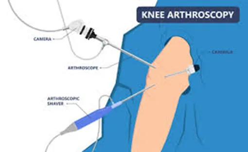
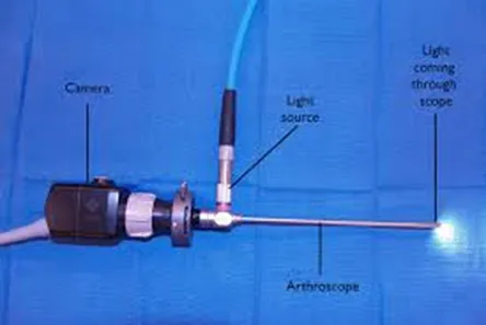
Knee arthroscopy (also known as keyhole knee surgery) is a minimally invasive procedure that uses a small camera, called an arthroscope, to view the inside of the knee joint.

This advanced technology enables surgeons to diagnose issues and perform treatments with precision through small incisions.

Using state-of-the-art tools and techniques, this procedure is designed to provide relief with minimal discomfort and downtime.

Whether you’re an athlete recovering from an injury or someone managing chronic knee pain, knee arthroscopy offers a solution tailored to your needs.

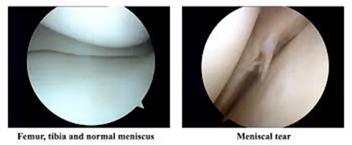
It’s commonly used for conditions such as meniscus tears, ligament injuries, and cartilage damage.

Who can benefit from knee arthroscopy?

This procedure is ideal for individuals who:

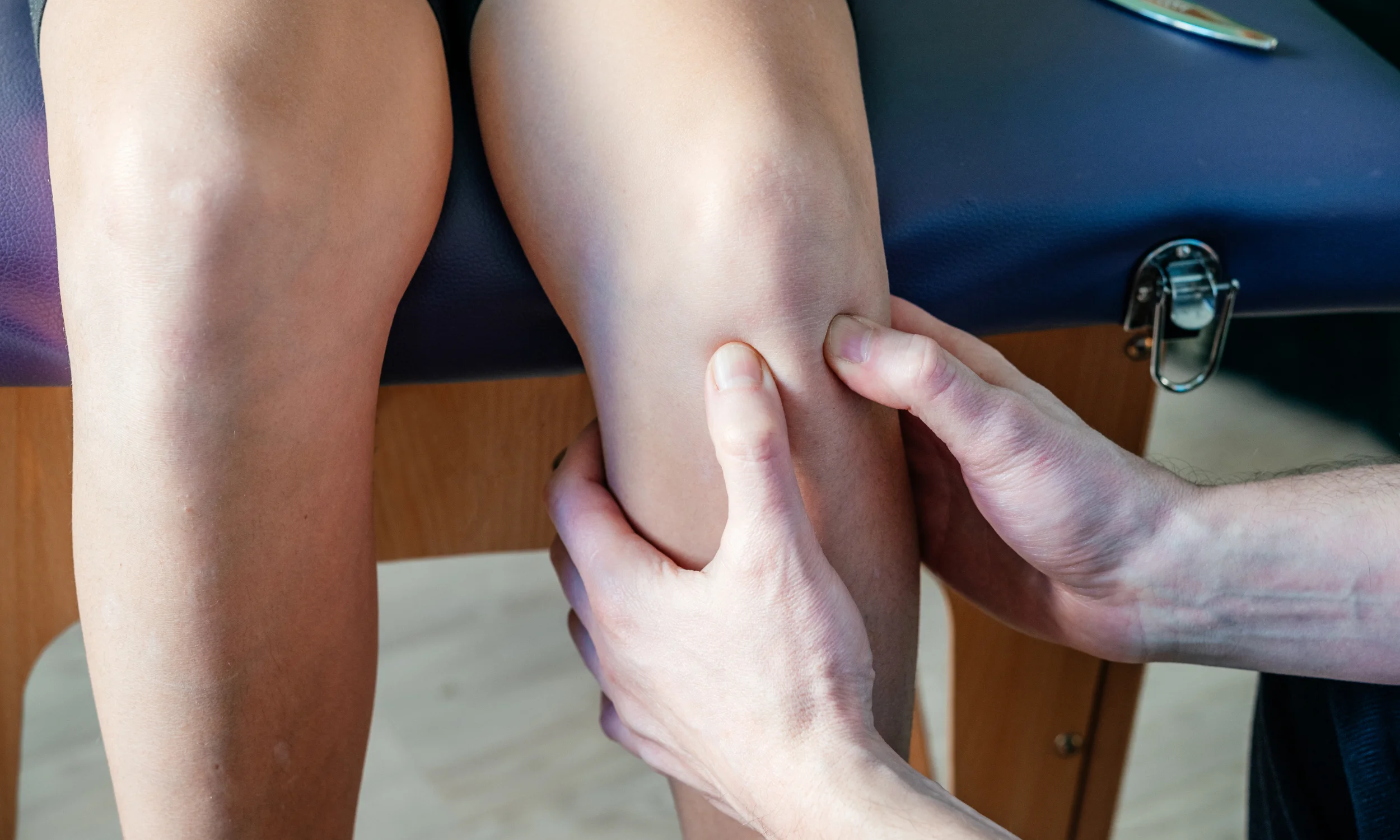
  • Have persistent knee pain that doesn’t improve with non-surgical treatments.
  • Experience limited range of motion or joint instability.
  • Have suffered injuries like torn ligaments or cartilage.
  • Need a definitive diagnosis of unexplained knee issues.

Knee arthroscopy is suitable for both active individuals and those dealing with degenerative conditions, offering a pathway to improved joint function and mobility.

What are the benefits of knee arthroscopy surgery?
  • Minimally invasive with small incisions and minimal scarring.
  • Reduced pain and faster recovery compared to open surgery.
  • High precision in diagnosing and treating knee conditions.
  • Ideal for athletes aiming for a quicker return to sports.

What are the common treatments performed during knee arthroscopy?

Knee arthroscopy can address various conditions and injuries using minimally invasive techniques.

Common treatments include:

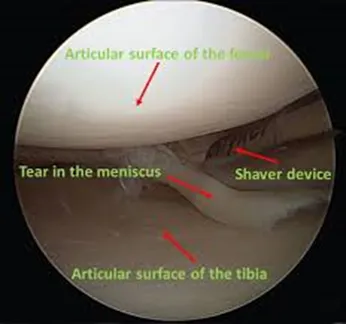
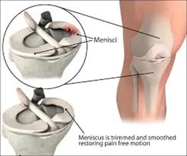
  • Meniscus tears: Procedures like meniscal repair or partial meniscectomy.
  • Cartilage injuries: Treatments such as microfracture or chondroplasty to restore or improve cartilage health.
  • Ligament reconstructions: Commonly performed for the anterior cruciate ligament (ACL).
  • Loose bodies removal:  This is where loose pieces of bone/cartilage are removed which are often fragments of cartilage from prior injuries.
  • Diagnostic assessment:  To assess the knee joint under direct vision using a camera.


During the procedure, the surgeon uses specialised instruments to treat the condition. The diagnosis is typically confirmed by thoroughly examining the joint with the arthroscope before proceeding with treatment.

How is Knee Arthroscopy Performed?

The procedure typically involves the following steps:

  • Preparation: The knee is cleaned, and anaesthesia is administered (commonly general anaesthesia).
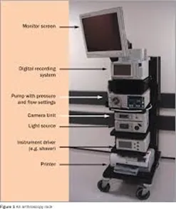
  • Arthroscope insertion: A small incision is made to insert the arthroscope, which transmits live images to a monitor.
  • Diagnosis and treatment: Using specialised instruments, the surgeon diagnoses the issue and performs the necessary repairs or treatments, such as trimming/repairing torn meniscus, or repairing torn ligaments.
  • Completion: The incisions are closed, and a bandage is applied.


Knee arthroscopy is often completed in less than an hour, depending on the complexity of the condition being treated.

What are the potential risks and complications?

Your surgeon takes every precaution to minimise risks of surgery, using state-of-the-art techniques and providing comprehensive preoperative and postoperative care.

Rest assured, your surgeon will carefully assess the risks and benefits before recommending surgery.

During your initial consultation, they will also discuss any specific risks relevant to your situation.

While knee arthroscopy is generally safe, some of the potential risks include:

  • Infection
  • Bleeding
  • Nerve injury
  • Stiffness or swelling
  • Scarring
  • Blood clots
  • Pain
  • Rare complications with anaesthesia


Our team prioritises safety and ensures every patient receives comprehensive care to minimise risks and promote optimal recovery.

What is the recovery time frames following surgery? 

The arthroscopy procedure causes minimal tissue damage, depending on the extent of the intervention undertaken. 

Most patients recover relatively quickly.

  • Hospital stay: Patients typically are discharged the same day following surgery. 
  • Pain management: Medications and physical therapy help manage postoperative pain.
  • Rehabilitation: Exercises to strengthen muscles and improve joint movement start shortly after surgery.


Time frames and post-op rehabilitation will depend on the exact treatment carried out during the surgery and should always be confirmed by your surgeon for individual cases.

As a general guide:

  • 1–2 days after surgery
    • You can eat and drink shortly after the procedure and should be able to walk, though crutches may be necessary for support.
    • A brace may be provided dependent on the nature of the surgery carried out during the procedure e.g. meniscal repair patient often provided with knee brace.
    • Avoid long walks or prolonged standing. When climbing stairs, lead with your good knee going up and your treated knee coming down.
  • 3 days after surgery
    • The surgeon will often recommend removing the wool and crepe bandage at 72 hours following the surgery, but to leave the ‘sticky wound dressings on’.  This needs to remain in situ till wound inspection by a health care professional usually at 2 weeks.  It is critically important to keep the wound dry during healing which can take up to 3 weeks and so protect it when having a shower.  Before leaving the hospital, your healthcare team will provide detailed instructions on wound care.
  • 1–2 Weeks after surgery
    • Some patients may still observe pain and swelling and should still avoid overexerting themselves. 
    • Elevation of the knee using a pillow under the foot at nighttime, and application of ice can help reduce swelling more quickly.
    • Physiotherapy exercises are important.
  • 2 Weeks after surgery 
    • A wound check is advised by a healthcare professional to ensure all has healed as expected.
    • The stitches from the keyhole sites if used are removed at this time.
  • 6–12 Weeks After Surgery
    • Most patients feel significantly recovered by six weeks, though some may need up to 12 weeks to feel fully normal.
    • Recovery time varies depending on your condition and the procedures performed.
    • Follow-up appointments with your surgeon at 6-8 weeks following surgery are important. During these visits:  The surgeon will assess wound healing, knee movement, and determine the need for additional physiotherapy.
  • Returning to Sports
    • Avoid sports until the swelling subsides and your knee is strong enough to handle exercise.
    • Your surgeon and physiotherapist will guide you on the appropriate timeline to resume sports safely dependent on the exact surgery carried out..
  • Returning to work 
    • Before going back to work, consult your surgeon for personalised advice as it will depend on the treatment carried out within the knee.
  • Returning to driving 
    • Refrain from driving until knee pain, swelling, and stiffness are resolved, and you regain full movement.
    • You may be able to drive again approximately six weeks after your surgery, although this timeframe can vary between individuals. Your consultant will provide specific guidance based on your progress.
    • Do not drive until you feel confident in controlling your vehicle and always confirm with both your doctor and your insurance provider before resuming driving.

What is the follow-up following surgery?
  • A wound check and removal of stitches (if used) will be required at approximately 14 days after your surgery with a healthcare professional.
  • A routine follow-up appointment with your surgeon will be needed around 6–8 weeks after your surgery.

How soon can you fly after knee arthroscopy surgery?

There is no specific formal medical guidance with respect to how long after surgery it might be before you are safe to fly.

Most surgeons and airlines recommend you should not fly within 6 weeks of surgery because of the increased risk of developing blood clots (Deep vein thrombosis, pulmonary embolism).

Please check with your surgeon for any advice regarding air travel and any precautions to take if you have additional risk factors for blood clots.


What is the recommended rehabilitation?  Tips for a smooth recovery

To strengthen your knee protect it from further injury, and support wound healing, follow these guidelines:

  • Perform the exercises recommended by your physiotherapist 3 to 4 times daily for 6 weeks. Listen to your body and do not worry if cannot keep up as recommended.
  • Elevation of the knee with a pillow under the foot (not under the knee) every nighttime over the first 4 weeks can help eventually reduce swelling more quickly.
  • If you experience discomfort, take the prescribed painkillers until the pain subsides.
  • It is critically important to keep the wound dry during healing which can take up to 3 weeks and so protect it when having a shower despite the dressing.


These steps will help ensure a safe and effective recovery.


How to maintain a healthy knee after surgery?
  • Follow rehabilitation guidelines: Adhere to physiotherapy and exercise routines. 
  • Prevent falls: Use assistive devices and take precautions to avoid accidents.
  • Healthy lifestyle: Maintain a balanced diet and healthy weight to reduce stress on the joint.
  • Regular check-ups: Monitor the condition of the prosthesis through routine medical evaluations.
Summary: Why choose knee arthroscopy?
  • A minimally invasive option for diagnosing and treating knee issues.
  • Ideal for addressing injuries and conditions that impact daily life or athletic performance.
  • Faster recovery and less discomfort compared to traditional surgery.
  • Performed by a skilled surgeon using advanced technology for precise results.


Take the first step towards better knee health. Schedule a consultation today to explore how knee arthroscopy can help you achieve pain-free mobility and improved joint function.

Physiotherapy

Knee physiotherapy protocols following knee replacement surgery

Physiotherapy is an essential part of recovery after knee replacement surgery. It helps restore mobility, improve strength, and ensure the best long-term outcomes. Below is a general outline of knee physiotherapy protocols, which may be tailored to each patient’s specific needs by their healthcare provider:

1. Immediate Postoperative Phase (Days 1–7)

Goals:

  • Prevent complications like blood clots or stiffness.
  • Begin gentle movement to restore range of motion.

Activities:

  • Ankle Pumps: To promote blood circulation.
  • Quad Sets: Tighten thigh muscles to prevent muscle atrophy.
  • Passive Range of Motion: Assisted bending and straightening of the knee.
  • Walking with Assistance: Using a walker or crutches for short distances.
2. Early Recovery Phase (Weeks 2–6)

Goals:

  • Increase range of motion (aim for 90–120 degrees of knee flexion).
  • Improve weight-bearing ability.
  • Strengthen surrounding muscles.

Activities:

  • Straight Leg Raises: Strengthen quadriceps without stressing the joint.
  • Heel Slides: Gradual bending of the knee while lying down.
  • Stationary Cycling: Low resistance to enhance mobility.
  • Step-Ups: Gentle step exercises on a low platform.
3. Intermediate Phase (Weeks 6–12)

Goals:

  • Normalize walking patterns.
  • Enhance muscle strength and joint stability.

Activities:

  • Balance Exercises: Use a stability board or single-leg stance.
  • Resistance Training: Light weights or resistance bands for leg muscles.
  • Walking Without Support: Gradually increase distances.
  • Functional Movements: Gentle squats and lunges to prepare for daily activities.
4. Advanced Phase (Months 3–6)

Goals:

  • Achieve full range of motion.
  • Return to low-impact activities.

Activities:

  • Higher Resistance Training: Focus on strengthening quads, hamstrings, and calves.
  • Advanced Balance Training: Progressively challenging exercises to improve coordination.
  • Low-Impact Activities: Swimming, walking, or cycling for cardiovascular fitness.
5. Maintenance Phase (Beyond 6 Months)

Goals:

  • Maintain strength and mobility.
  • Safely return to routine activities or sports, as advised.

Activities:

  • Continue with strengthening and mobility exercises.
  • Low-impact sports and recreational activities.
  • Periodic follow-ups with a physiotherapist for guidance.
Key Points to Remember:
  • Consistency is crucial for optimal recovery.
  • Avoid high-impact activities that may stress the joint.
  • Always follow your surgeon’s and physiotherapist’s advice regarding activity progression.
Hip physiotherapy protocols following hip replacement surgery

Physiotherapy after hip replacement surgery plays a vital role in regaining mobility, strengthening muscles, and ensuring the longevity of the new joint. Below is an overview of the typical physiotherapy protocols, divided into phases for a structured recovery process:

1. Immediate Postoperative Phase (Days 1–7)

Goals:

  • Prevent complications like blood clots or stiffness.
  • Begin gentle movement and weight-bearing exercises.

Activities:

  • Ankle Pumps: To improve blood circulation and reduce swelling.
  • Quad Sets and Glute Squeezes: Strengthen thigh and hip muscles.
  • Heel Slides: Gradual bending of the hip and knee while lying down.
  • Assisted Walking: Use of a walker or crutches for short distances.
2. Early Recovery Phase (Weeks 2–6)

Goals:

  • Improve range of motion and muscle strength.
  • Transition to more independent mobility.

Activities:

  • Straight Leg Raises: Strengthen quadriceps without stressing the hip.
  • Hip Abductions: Lifting the leg sideways while lying down or standing.
  • Stationary Cycling: Gentle cycling with low resistance.
  • Weight-Bearing Exercises: Gradual increase in walking distances with support.
3. Intermediate Recovery Phase (Weeks 6–12)

Goals:

  • Normalize walking patterns and restore functional movement.
  • Strengthen surrounding muscles and improve joint stability.

Activities:

  • Step-Ups: Controlled step exercises on a low platform.
  • Balance Training: Single-leg stance exercises with support if needed.
  • Resistance Band Workouts: Strengthen hip abductors and extensors.
  • Walking Without Support: Gradually increase distances, focusing on proper gait.
4. Advanced Recovery Phase (Months 3–6)

Goals:

  • Build strength and flexibility to support routine activities.
  • Begin low-impact recreational activities.

Activities:

  • Advanced Strengthening Exercises: Light weights for leg muscles.
  • Functional Training: Gentle squats and lunges to prepare for daily activities.
  • Low-Impact Aerobic Activities: Swimming, cycling, or walking for endurance.
5. Maintenance Phase (Beyond 6 Months)

Goals:

  • Maintain strength and mobility.
  • Safely resume hobbies and low-impact sports.

Activities:

  • Continue strengthening exercises and stretching routines.
  • Engage in low-impact activities like yoga or Pilates.
  • Follow up with your physiotherapist periodically to ensure progress.
Important Tips:
  • Avoid high-impact activities or heavy lifting during the early phases.
  • Adhere to hip precautions (e.g., avoiding excessive bending or twisting) as advised by your surgeon.
  • Pain is a signal to stop—don’t push through discomfort.
  • Stay consistent with your physiotherapy exercises for optimal recovery.
Knee physiotherapy protocols following knee meniscal surgery (meniscus repaired vs trimmed (partial meniscectomy)
1. Post-Operative Phase: (Weeks 1-2)

Goal: Control pain, swelling, and inflammation, protect the surgical site, and initiate early range of motion (ROM).

  • Meniscal Repair Protocol:
    • Weight-bearing: Non-weight bearing (NWB) or partial weight-bearing (PWB) with crutches for 4-6 weeks to protect the repair.
    • ROM: Passive and active-assisted range of motion exercises within pain tolerance.
    • Exercise: Isometric quadriceps contractions, ankle pumps, and straight leg raises to avoid muscle atrophy.
    • Protection: Knee brace with limited flexion (0-90 degrees), as directed by the surgeon.
  • Partial Meniscectomy Protocol:
    • Weight-bearing: Full weight-bearing (FWB) allowed immediately or after a few days depending on the surgeon's preference.
    • ROM: Gentle range of motion exercises, avoiding deep squats or high-load activities.
    • Exercise: Focus on quadriceps strengthening, and gentle hamstring and calf stretches.
    • Protection: No bracing is generally needed, but the patient should avoid twisting or pivoting.
2. Early Rehabilitation Phase: (Weeks 3-6)

Goal: Improve ROM, decrease swelling, and initiate strength training.

  • Meniscal Repair Protocol:
    • Weight-bearing: Gradual increase in weight-bearing, progressing to full weight-bearing by 6 weeks if tolerated.
    • ROM: Continue working on achieving full extension and 90-110 degrees of flexion.
    • Strengthening: Quadriceps and hamstring strengthening exercises, focusing on pain-free motion.
    • Exercise: Progress to closed-chain exercises like leg presses (avoiding deep knee bends initially), hip abduction, and mini squats.
  • Partial Meniscectomy Protocol:
    • Weight-bearing: Full weight-bearing is maintained.
    • ROM: Full knee extension and flexion should be encouraged.
    • Strengthening: Quadriceps and hamstring strengthening with light resistance bands or weights.
    • Exercise: Begin low-impact closed-chain exercises, including stationary cycling and pool therapy if available.
3. Intermediate Rehabilitation Phase: (Weeks 7-12)

Goal: Build strength, improve functional mobility, and gradually return to more dynamic activities.

  • Meniscal Repair Protocol:
    • Strengthening: Progressive resistance training (leg press, squats) and proprioceptive exercises (e.g., balance and stability work).
    • Activity: Begin low-impact activities such as cycling and swimming, avoid running or jumping.
    • Functional Training: Begin agility drills and proprioceptive training.
  • Partial Meniscectomy Protocol:
    • Strengthening: Continue strengthening exercises with added resistance.
    • Activity: Running can begin if there are no significant signs of pain or swelling.
    • Functional Training: Return to light activities, with controlled motion, agility drills, and functional tasks.
4. Advanced Rehabilitation Phase: (Weeks 13-24)

Goal: Continue to strengthen the knee, return to normal function, and resume sports and recreational activities.

  • Meniscal Repair Protocol:
    • Strengthening: Continue strengthening exercises (focus on quads, hamstrings, and calves) and increase resistance or weights as tolerated.
    • Activity: Low-impact activities like swimming or cycling. Running may be introduced cautiously at 12-16 weeks, depending on the surgeon's approval.
    • Return to Sports: Typically after 6 months, depending on healing progress, sports-specific exercises may begin.
  • Partial Meniscectomy Protocol:
    • Strengthening: Full return to weight-bearing exercises with more advanced resistance.
    • Activity: Gradual progression of running, plyometrics, and sport-specific drills.
    • Return to Sports: Return to full activity and sports after 12-16 weeks if cleared by the surgeon.
5. Long-Term Maintenance Phase: (Months 6+)

Goal: Maintain knee strength and function and prevent further injury.

  • Meniscal Repair Protocol:
    • Full return to sports may take 6-12 months or more, depending on the type and success of the meniscal repair.
    • Continued strengthening and conditioning exercises.
  • Partial Meniscectomy Protocol:
    • Return to normal activity should be well-established by 6 months.
    • Ongoing strengthening exercises and proprioceptive training to ensure knee stability.
Key Differences:
  • Weight-bearing restrictions: More stringent in meniscal repair versus partial meniscectomy.
  • Return to activity: Generally, patients following meniscal repair take longer to return to sports due to the need for healing of the repaired meniscus.
  • Intensity of exercise: Those with a meniscal repair often progress more slowly in terms of weight-bearing and exercise intensity.

It is crucial for patients to follow these protocols closely, with regular follow-up appointments with their surgeon or physiotherapist to ensure proper healing and recovery progress.

Physiotherapy to manage hip pain from osteoarthritis conservatively

Managing hip pain from osteoarthritis (OA) conservatively through physiotherapy involves a combination of exercises, manual therapy, and lifestyle modifications aimed at reducing pain, improving function, and preventing further deterioration of the joint. Here's a detailed approach to physiotherapy for managing hip pain caused by OA

1.Pain Management
  • Heat/Cold Therapy: Use cold packs to reduce inflammation and heat to relax muscles.
  • TENS Therapy: Electrical stimulation can help manage pain.
2. Strengthening Exercises
  • Glutes and Quads: Exercises like leg raises, squats, and bridges help stabilize the hip joint.
  • Hamstrings: Strengthening the hamstrings to balance leg muscles and reduce stress on the hip.
3. Range of Motion (ROM) Exercises
  • Hip Flexion/Extension Stretching: Gentle stretches to improve flexibility and reduce stiffness.
  • Rotator Stretching: Helps improve internal and external hip rotation.
4. Manual Therapy
  • Joint Mobilization: Gentle, hands-on techniques to improve joint movement.
  • Soft Tissue Mobilization: Focus on relieving tightness in surrounding muscles like the hip flexors and glutes.
5. Posture and Gait Training
  • Posture Awareness: Educate on proper body mechanics to avoid excess strain on the hip joint.
  • Gait Training: Adjust walking techniques to reduce impact on the hip.
6. Core Stability and Balance
  • Core Strengthening: Exercises like planks to improve posture and reduce hip strain.
  • Balance Training: Simple exercises like single-leg stands to improve stability.
7. Aquatic Therapy
  • Water Exercises: Buoyancy reduces joint stress, making it easier to perform strengthening and stretching exercises.
8. Long-Term Maintenance
  • Home Exercise Program: Provide simple exercises for patients to do at home.
  • Gradual Activity Increase: As strength and mobility improve, encourage more activity like swimming and walking.
physiotherapy to manage hip pain from trochanteric hip bursitis

Trochanteric hip bursitis causes pain on the outer side of the hip due to inflammation of the bursa. Physiotherapy can help reduce pain, improve function, and prevent recurrence.

1. Pain Management
  • Ice Therapy: Apply ice to the outer hip for 15-20 minutes, several times a day to reduce inflammation and pain.
  • Heat Therapy: Use heat to relax tight muscles once acute pain subsides.
  • TENS Therapy: Electrical nerve stimulation can assist with pain relief.
2. Stretching Exercises
  • Hip Flexor Stretch: Relieves tightness in the front of the hip.
  • IT Band Stretch: Reduces tension on the outer hip.
  • Glute Stretch: Helps ease tightness in the glutes and surrounding muscles.
3. Strengthening Exercises
  • Gluteus Medius Strengthening: Side-lying leg lifts and clamshells target the gluteus medius to support the hip.
  • Hip Abduction: Strengthens the hip abductors to stabilize the joint and reduce stress on the bursa.
  • Core Strengthening: Bridges and planks improve pelvic stability and reduce strain on the hip.
4. Manual Therapy
  • Soft Tissue Mobilization: Gentle massage can relieve muscle tightness around the hip.
  • Joint Mobilizations: Physiotherapists may use manual techniques to enhance hip mobility.
5. Posture and Gait Training
  • Postural Awareness: Correct standing, sitting, and walking positions reduce hip strain.
  • Gait Training: Proper walking mechanics can prevent aggravation of the bursitis.
6. Balance and Stability Training
  • Balance Exercises: Single-leg stands improve stability and reduce compensatory movements.
  • Functional Exercises: Gradually incorporate exercises that mimic daily activities to strengthen the hip.
7. Gradual Return to Activity
  • Low-Impact Activities: Start with swimming or cycling to strengthen without stressing the hip.
  • Avoid Overuse: Gradually increase activity levels to prevent flare-ups.

This conservative approach can significantly reduce pain and improve hip function in individuals with trochanteric hip bursitis.

Physiotherapy to manage knee pain from osteoarthritis conservatively

Physiotherapy plays a crucial role in managing knee pain from osteoarthritis (OA) conservatively by focusing on pain relief, improving joint function, and preventing further damage. Here's a concise breakdown of the approach:

1. Pain Management
  • Ice Therapy: Apply ice to the knee for 15-20 minutes to reduce inflammation and pain.
  • Heat Therapy: Use heat to relax muscles around the knee after acute pain subsides.
  • TENS Therapy: Transcutaneous electrical nerve stimulation can provide pain relief by stimulating nerves.
2. Strengthening Exercises
  • Quadriceps Strengthening: Exercises like straight leg raises and squats help strengthen the quadriceps, which support the knee joint.
  • Hamstring Strengthening: Strengthening the hamstrings reduces strain on the knee and balances the muscles around the joint.
  • Glute Strengthening: Targeting the glutes with exercises like bridges helps improve pelvic stability and reduce knee strain.
3. Range of Motion (ROM) Exercises
  • Knee Flexion/Extension: Gentle stretches to improve the knee's ability to bend and straighten.
  • Heel and Toe Raises: To maintain mobility and reduce stiffness in the knee joint.
4. Manual Therapy
  • Joint Mobilization: Hands-on techniques from the physiotherapist can improve knee mobility and alleviate discomfort.
  • Soft Tissue Mobilization: Massage or myofascial release to ease muscle tightness and improve blood flow around the knee.
5. Posture and Gait Training
  • Postural Awareness: Correct alignment when standing, sitting, and walking can help reduce stress on the knee.
  • Gait Training: Learning to walk with a more efficient stride can alleviate pressure on the knee joint.
6. Balance and Stability Training
  • Balance Exercises: Single-leg stands or balance boards can improve knee stability and prevent falls.
  • Proprioception Training: Exercises that help improve the knee’s sense of position can reduce the risk of injury.
7. Aquatic Therapy
  • Water-Based Exercises: Swimming or water aerobics can reduce impact on the knee while strengthening muscles and improving range of motion.
8. Gradual Return to Activity
  • Low-Impact Activities: Activities like cycling or walking can be introduced to build strength without overloading the knee.
  • Avoid Overuse: Gradually increasing activity levels ensures the knee is not overstrained.

This conservative physiotherapy approach aims to improve knee function, reduce pain, and help patients manage osteoarthritis effectively.